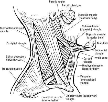
Medially sagittal line down the midline of the neck.
Roof of digastric triangle.
The digastric triangle is one of the paired triangles in the anterior triangle of the neck.
The digastric triangle is subdivided into anterior and posterior parts by the stylomandibular ligament which goes from the tip of the styloid process to the angle of the mandible.
The triangles of the neck are surgically focussed first described from early dissection based anatomical.
The posterior part of the triangle is constant above with the parotid region.
The submandibular triangle or submaxillary or digastric triangle corresponds to the region of the neck immediately beneath the body of the mandible boundaries and coverings.
This triangle contains major arteries veins and nerves of the neck and head.
The anterior triangle is situated at the front of the neck.
Floor posteriorly is inferior pharyngeal conatrictor muscle anteriorly is the thyrohyoid muscle and hypoglossus roof is investing layer of fascia cutaneous nerves and platysma.
Anteriorly by the anterior belly of digastric muscle.
Its roof is formed by deep and superficial fascia platysma and skin.
The branches of the facial nerve and transverse cutaneous cervical nerves also pass over the roof of the triangle.
Contents in the anterior part of the triangle.
Platysma cervical division of facial nerve and ascending branch of transverse cervical nerve are located in the superficial fascia above the roof.
The stylomandibular ligament subdivides the digastric triangle within anterior and posterior parts.
The roof of the triangle is formed the skin superficial cervical fascia the platysma and deep cervical fascia.
The digastric triangle is one of the paired triangles in the anterior triangle of the neck.
Above by the lower border of the body of the mandible and a line drawn from its angle to the.
Posterior belly of digastric muscle pbd superior belly of the omohyoid muscle so anterior border of sternomastoid muscle st roof.
The triangles of the neck are surgically focused first described from early dissection based anatomical studies which predated cross sectional anatomical description based on imaging see deep spaces of the neck.
Superiorly inferior border of the mandible jawbone.
Laterally anterior border of the sternocleidomastoid.
Posterior belly of digastric muscle posterior anterior portion of scm.
Skin superficial fascia platysma 17.
The posterior portion of the triangle is superiorly constant with the parotid region.
Investing fascia covers the roof of.

